Skip to content
STEMI
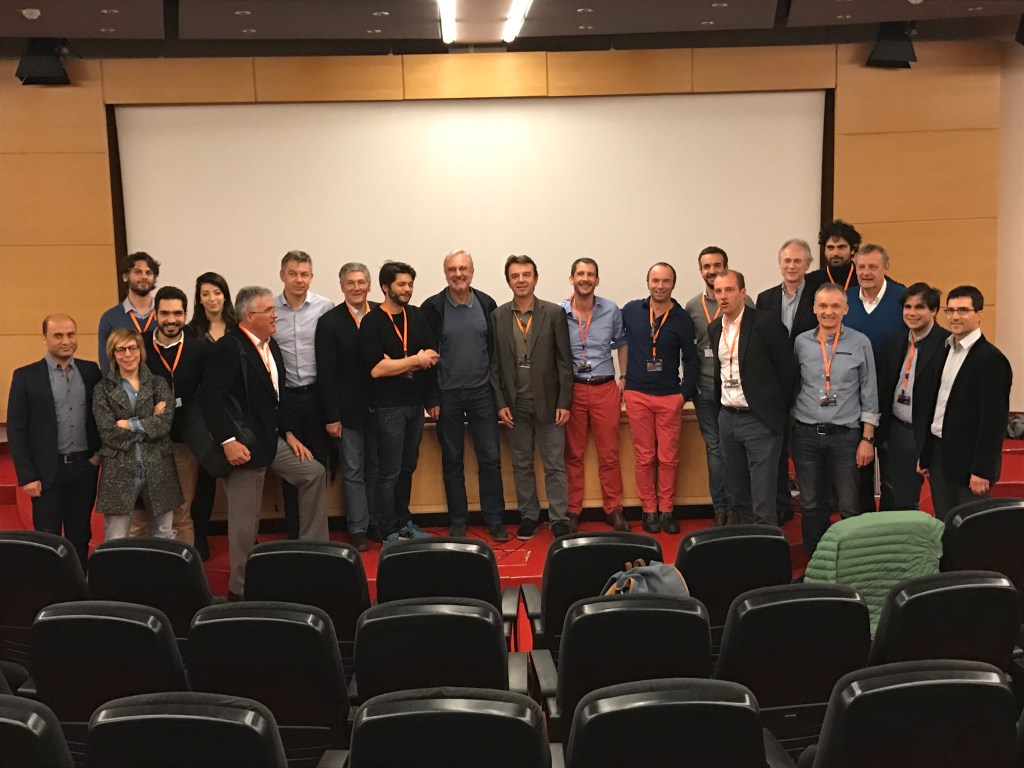
 A specialized medical team performs a complex heart procedure using real-time diagnostic imaging.
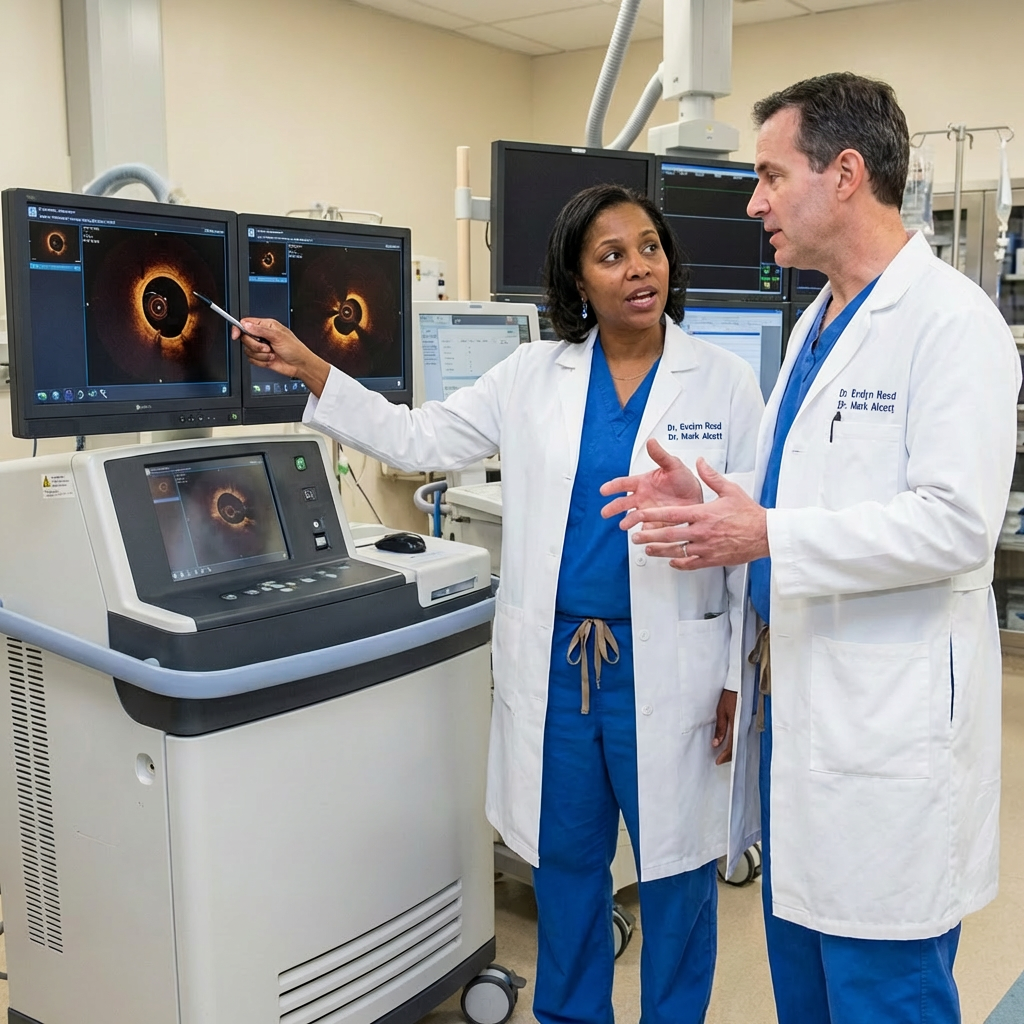 Medical professionals collaborate to analyze detailed cardiac imaging results in a clinical setting.
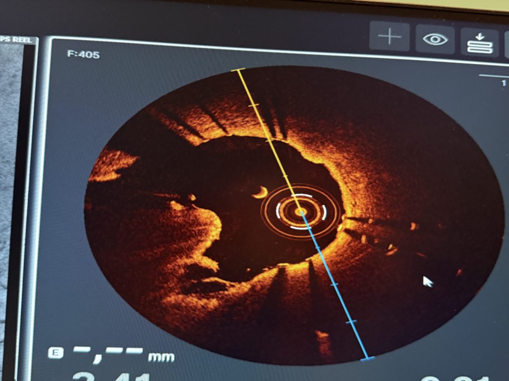
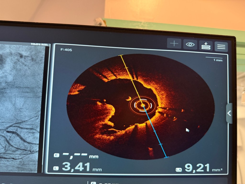
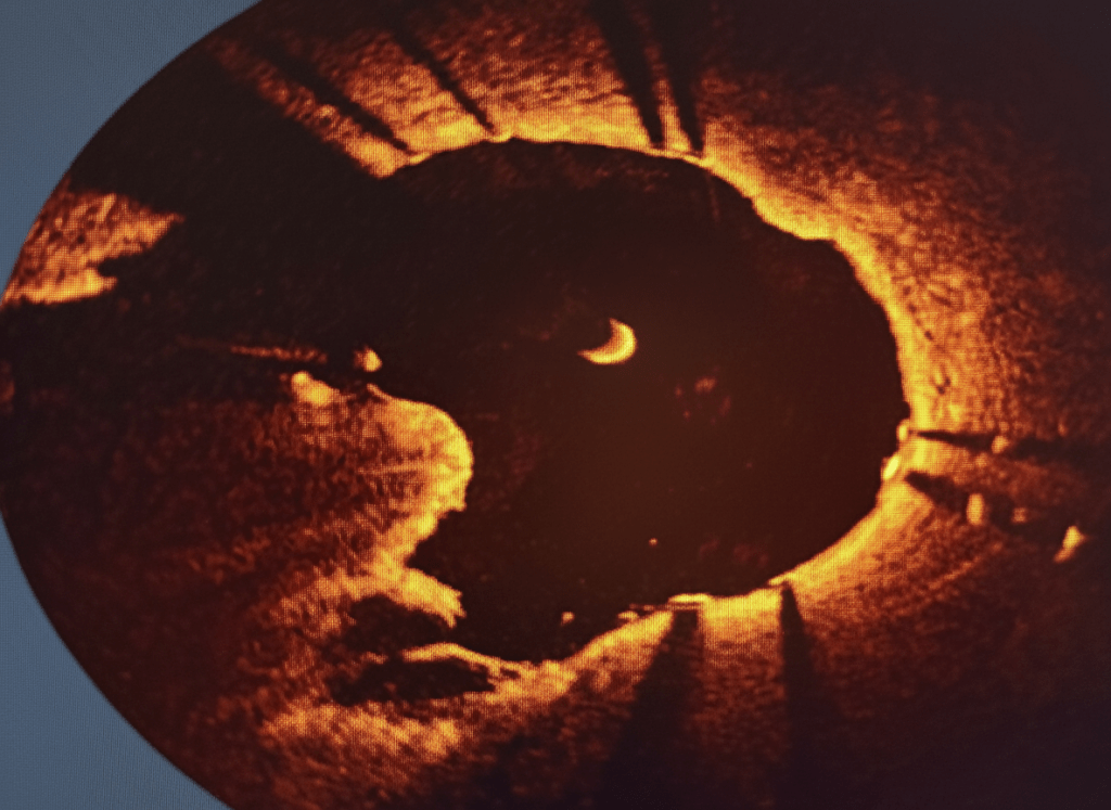
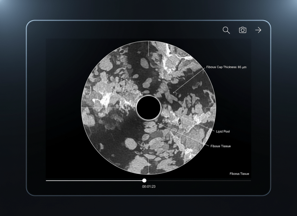
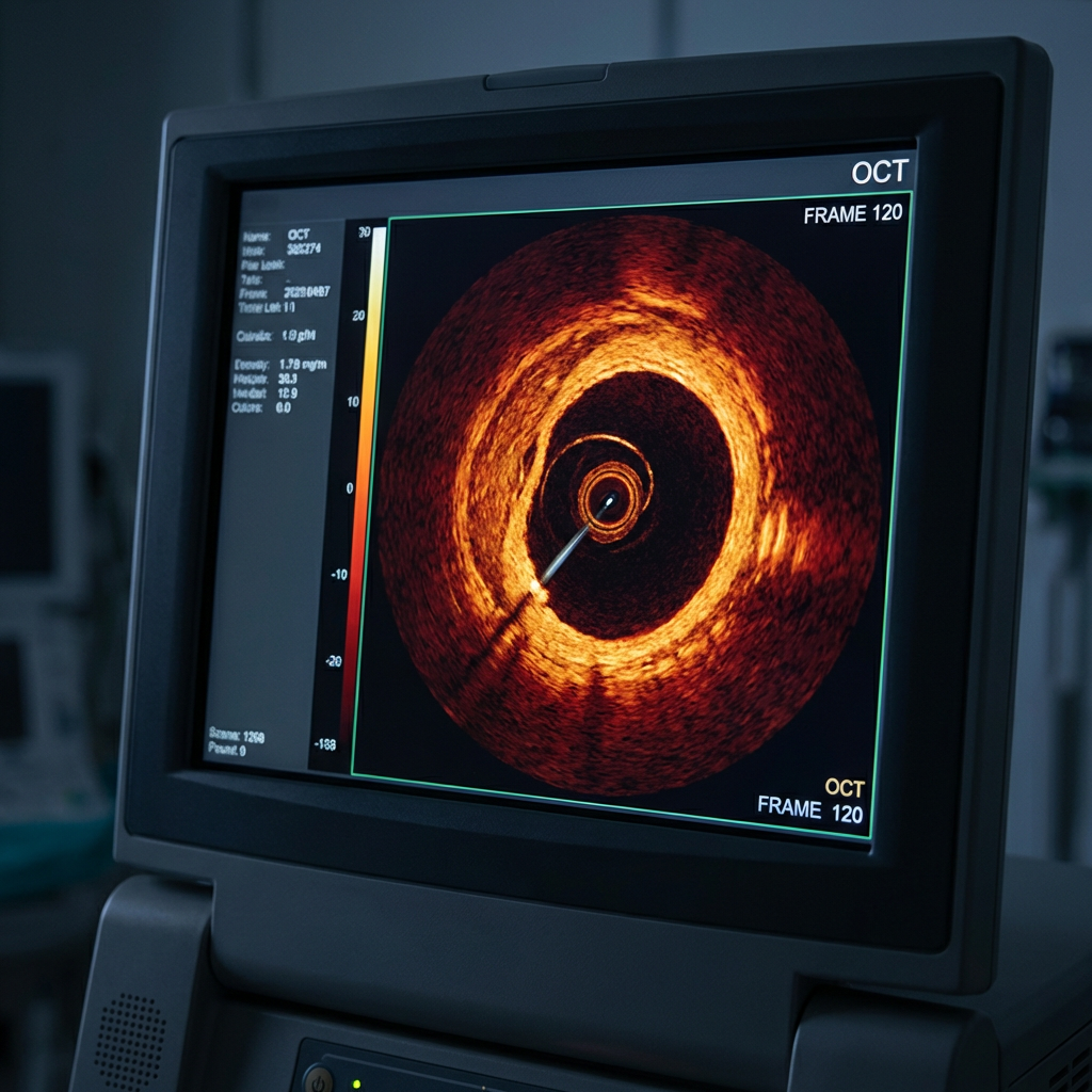 A specialized medical monitor displays a detailed cross-sectional scan of a blood vessel using OCT technology.
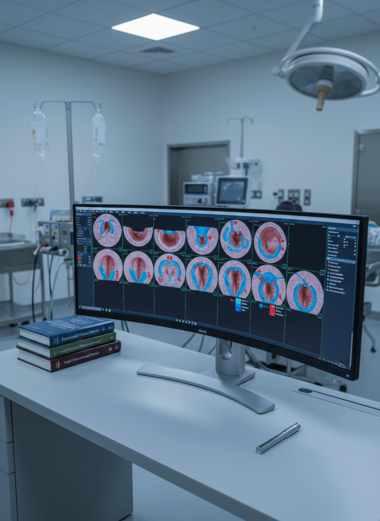
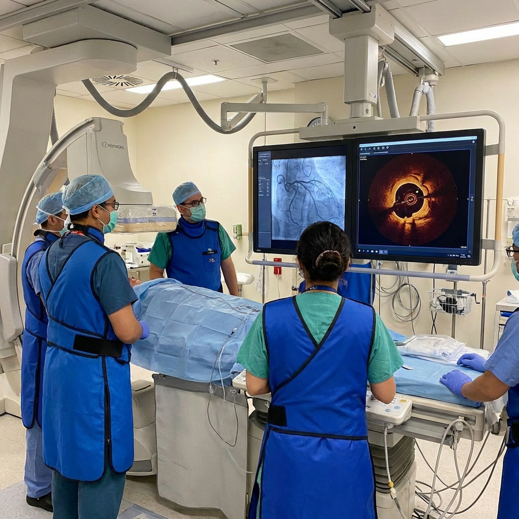
- A specialized medical team performs a complex heart procedure using real-time diagnostic imaging.
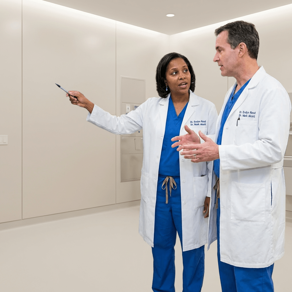
- Medical professionals collaborate to analyze detailed cardiac imaging results in a clinical setting.
- A specialized medical monitor displays a detailed cross-sectional scan of a blood vessel using OCT technology.
SCC
- A specialized medical team performs a complex heart procedure using real-time diagnostic imaging.
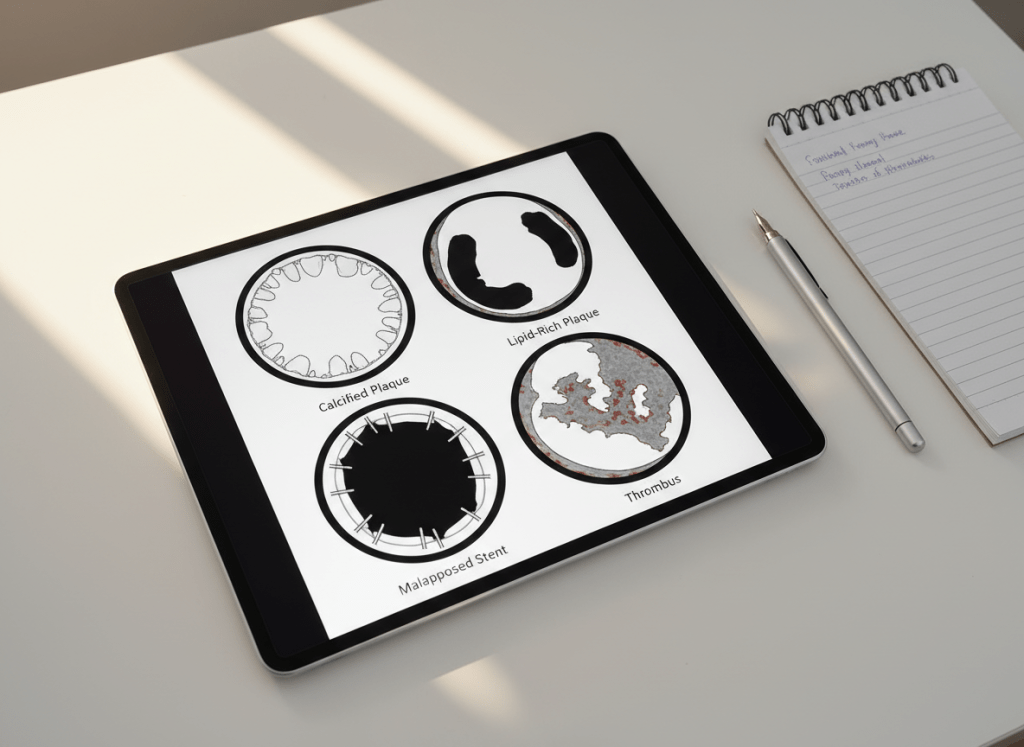
Calcified lesion
Ambigous morphology
SCAD
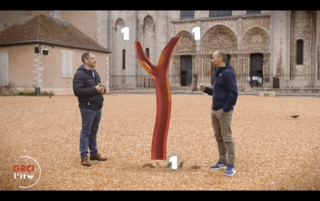
 left main