Case Library

Start with lesion type or stent status, then dive into annotated OCT cases and slides.

A detailed, photographic-style workstation setup featuring a large, ultra-wide medical-grade monitor displaying a vivid coronary OCT pullback sequence, with multiple frames aligned in series and color-coded annotations near areas of plaque. The monitor rests on a clean, light gray desk beside a neatly stacked set of cardiology textbooks and a slim, metallic digital caliper. Cool, diffused overhead lighting creates minimal glare, emphasizing clarity on the screen. The background is softly blurred, hinting at a modern catheterization lab environment with stainless steel equipment. Captured from a slightly elevated angle using a moderate depth of field, the image feels organized, analytical, and ideal for clinical education.
An educational, photographic-style layout of multiple coronary OCT frames arranged in a grid on a digital tablet screen, each frame illustrating a different pathology: calcified plaque with bright, sharply delineated nodules; lipid-rich plaque with signal-poor regions; malapposed stent struts; and thrombus with irregular protrusions into the lumen. The tablet rests on a smooth white surface beside a neatly arranged notepad and a precision pen. Soft, natural daylight from an unseen window casts gentle, diagonal shadows, creating a calm, studious atmosphere. Shot from a slightly elevated, top-down perspective with sharp focus, the composition is clean, modern, and tailored to an atlas-style educational resource.

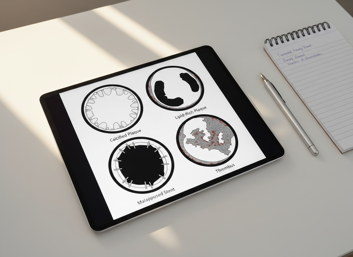
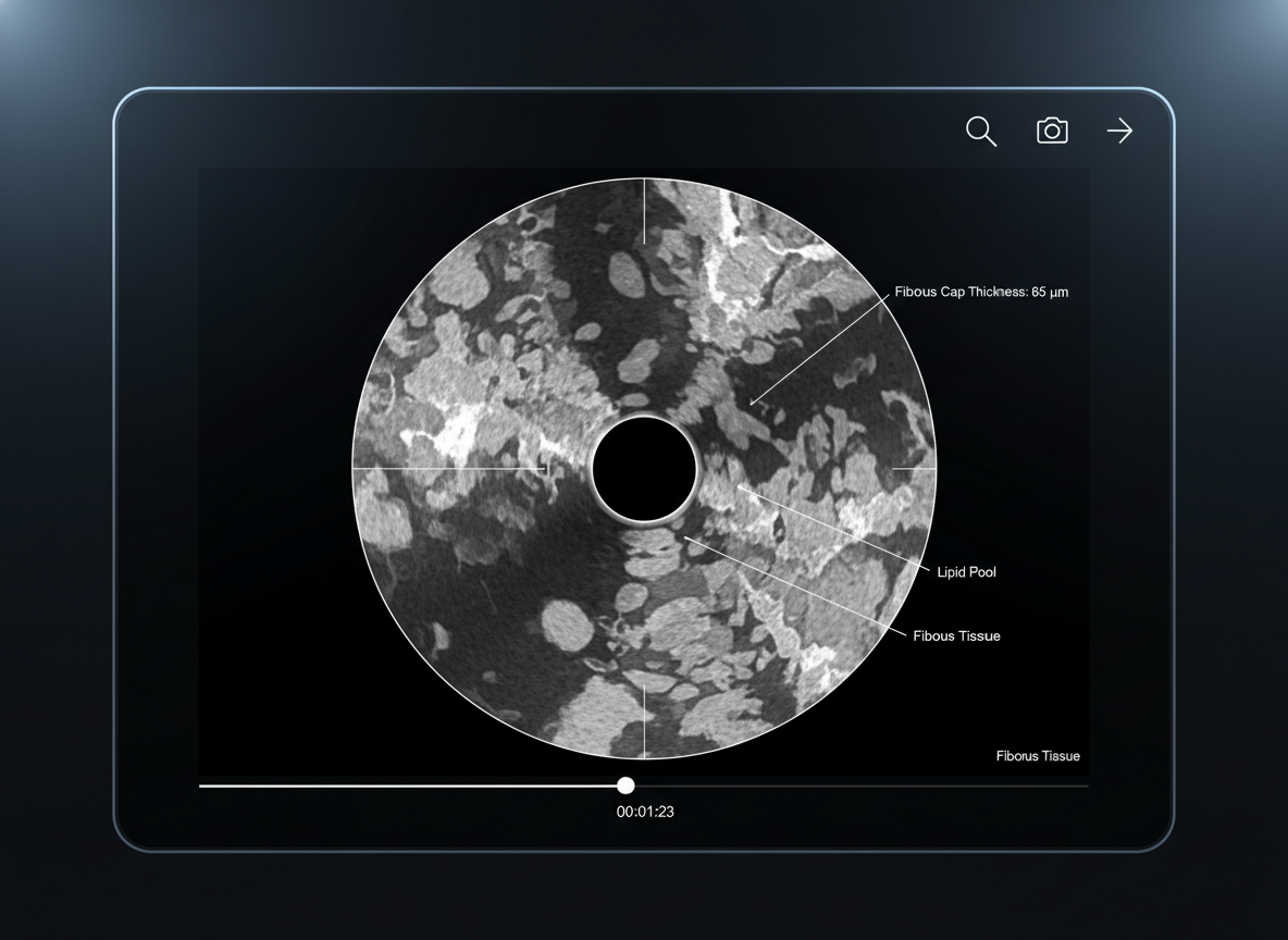
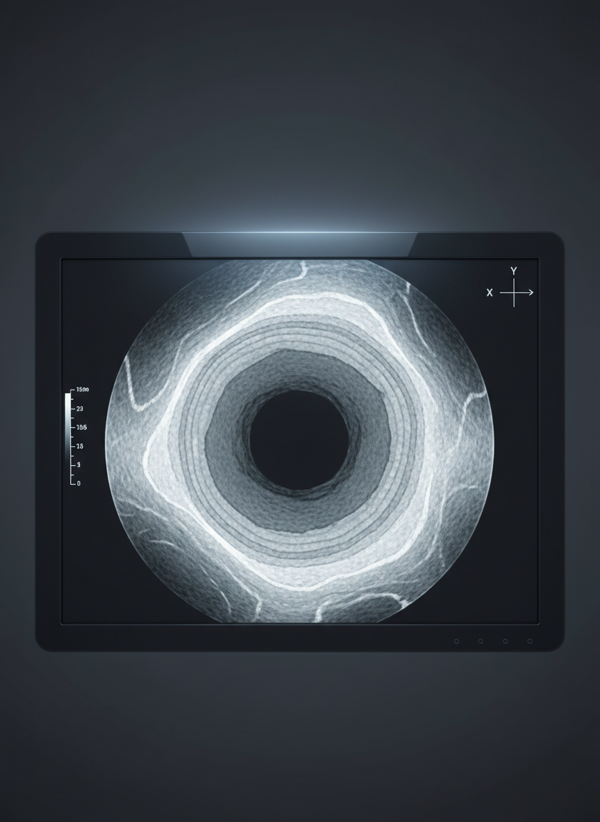
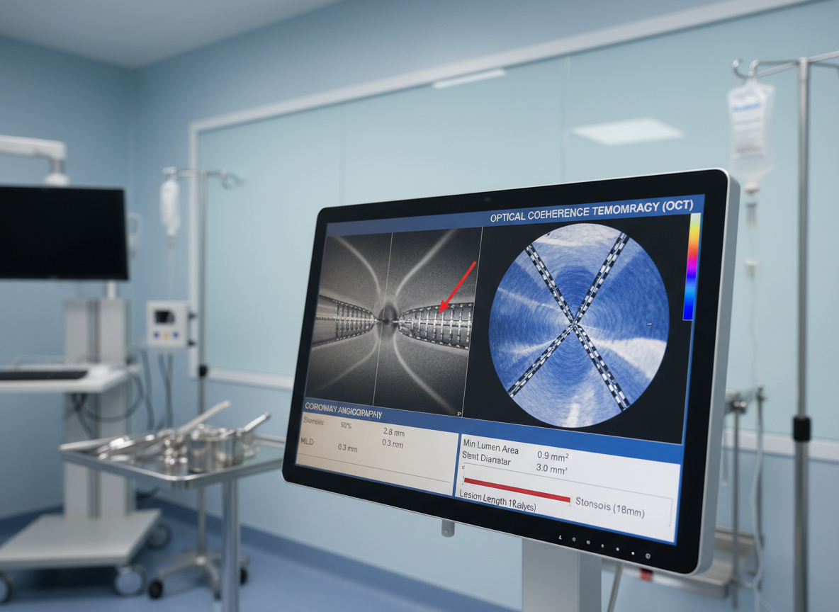
Coronary OCT Atlas Navigator

Browse our image atlas by lesion type, stent scenario, and common artifacts to quickly find comparable cases. Each category links to curated OCT pullbacks, expert interpretations, and teaching slides designed for everyday clinical decision‑making.

A close-up, photographic OCT image of a coronary artery with mixed atherosclerotic plaque, showing irregular, heterogeneous regions with varying backscatter and shadowing, contrasted against a smooth, circular lumen. Thin measurement lines and small labels indicate fibrous cap thickness and plaque components. The OCT frame is presented on a matte black digital viewer interface with minimalist icons and a discrete timeline bar at the bottom. Subtle, cool-toned studio lighting highlights the edges of the screen, creating a focused, high-tech mood. Shot in centered composition with sharp detail, the atmosphere is precise, didactic, and suited for explaining plaque morphology to clinicians.
A high-resolution, photographic cross-sectional view of a coronary artery rendered as a precise OCT image, showing concentric vessel layers with subtle gradations of gray and white, and clearly delineated lumen. The artery segment is displayed on a sleek, dark charcoal background with clean axis markers and small, unobtrusive measurement calipers along one edge. Soft, cool studio lighting illuminates the display, creating gentle reflections on the glossy monitor frame while keeping the image content crisp and legible. Shot straight-on at eye level with sharp focus throughout, the composition is centered and balanced, evoking a professional, clinical atmosphere suitable for an educational cardiology website.
A photographic depiction of a modern catheterization lab console screen displaying a side-by-side comparison of coronary angiography and OCT images of the same lesion, with the OCT frame showing a tight stenosis and clear stent struts. The interface includes neatly aligned analysis panels, numerical measurements, and a lesion length graph. The console is set against a softly lit, light blue clinical wall, with metallic instrumentation softly blurred in the background. Neutral, diffused overhead lighting ensures no harsh reflections, maintaining crisp legibility. Captured at a three-quarter angle with moderate depth of field, the composition suggests an advanced, data-driven environment ideal for teaching practical OCT interpretation.